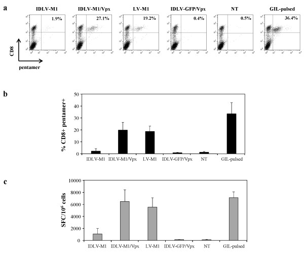
Figure 3.
Evaluation of M1-specific CD8+ T cell expansion by using transduced DC as antigen presenting cells. DC were transduced with IDLV-M1, IDLV-M1/Vpx, integrating LV-M1 or IDLV-GFP/Vpx as an unrelated antigen (MOI 1). M1-specific peptide-pulsed (GIL-pulsed) or unpulsed DC (NT) were used as positive and negative controls, respectively. For pentamer assay (a and b), cells were stained with anti-CD8 PE-Cy5 antibody and PE-labeled HLA-A*0201 pentamer presenting the influenza matrix M1 epitope. The percentage of pentamer + cells was calculated within CD8+ T cells and in (a) a representative experiment is shown. (b) Percentages of CD8+ pentamer + T cells evaluated in four different healthy donors are indicated. Graphs show means ± SD. (c) The functionality of expanded CD8+ T cells was evaluated by IFN-γ-ELISPOT in the presence of M1 specific peptide. Results are expressed as spot forming cells (SFC) per 1x10⁶ cells.

