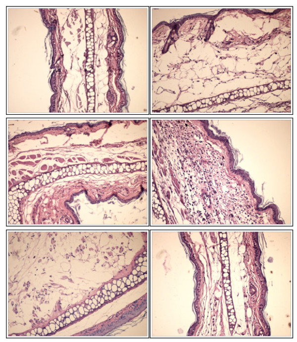
Figure 7.
Histological analysis. a) Normal tissue - Group A (HE×200); b) Massive oedema of the superficial and deep dermis. Major destruction of the superficial dermis. Complete lyses of the muscular layer- Group B (HE×200); c) Massive oedema of the deep dermis. Partial destruction of the superficial dermis. Partial destruction of the underlying muscle cells- Group C (HE×200); d) Rich polymorphous inflammatory infiltrate that increases in thickness the sub epithelial dermis without inflammatory infiltrate in epithelial tissue-Group D (HE×200); e) Epithelial and cartilaginous tissue with marked edema Group E (HE×200); f) Loose stroma, oedematous, with low cellularity-Group F (HE×200).

