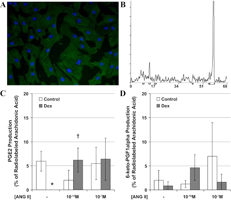
Fig. 2.
Second passage coronary myocytes obtained from adolescent sheep uniformly stained positive for α-smooth muscle actin (A, Dex-exposed cells with DAPI nuclear counterstain). Ninety minutes after incubation with radiolabeled arachidonic acid in the presence and absence of ANG II, lipids were extracted from the media, and eicosanoids were separated by HPLC with peaks identified for 6-keto-PGF1α (retention time 9 min), PGE2 (retention time 13 min), and arachidonic acid (retention time of 57 min) (B, representative run from control cells, y-axis: counts per minute, x-axis: retention time in minutes). The production of PGE2 (C) and 6-keto-PGF1α (D) was compared between control (open bars) and Dex-exposed sheep (solid bars). N = 4 sheep per group, *P < 0.05 vs. control, †P < 0.05 vs. buffer alone.

