Fig. 1.
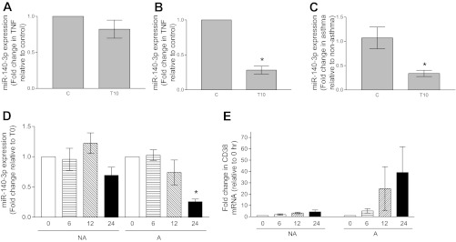
Expression of miR-140-3p in vehicle-treated (C) and TNF-α-treated (T10) nonasthmatic airway smooth muscle (NAASM) and asthmatic airway smooth muscle (AASM) cells. A: in the presence of TNF-α, miR-140-3p expression was marginally attenuated in NAASM cells compared with cells treated with vehicle (n = 5). B: in AASM cells, TNF-α significantly attenuated miR-140-3p expression (n = 6). *P < 0.05. C: basal miR-140-3p expression levels were comparable between NAASM and AASM cells. In the presence of TNF-α, miR-140-3p expression was significantly attenuated in AASM cells compared with NAASM cells. *P < 0.05. (Data in A–C are from the same experiments.) D: when NAASM (NA) and AASM (A) cells were exposed to TNF-α for 0–24 h, both showed attenuated miR-140-3p expression at 24 h, although reduction was statistically significant only in AASM cells (n = 3). *P < 0.05. E: TNF-α caused a time-dependent increase in CD38 mRNA expression in NAASM and AASM cells. Magnitude of CD38 induction in AASM cells was higher in AASM cells, although differential increase was not statistically significant because of larger standard error in AASM cells.

