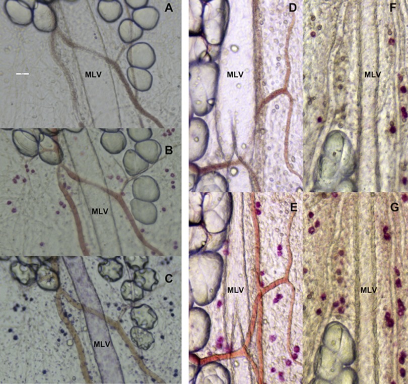
Fig. 2.
Representative images of the activation of mast cells located in close proximity to rat mesenteric lymphatic vessels in adult and aged segments of mesentery. A: segment (9 mo old) of mesentery before activation. B: same segment with mast cells activated by compound 48/80 stained with the ruthenium red. C: same segment stained with toluidine blue. D: segment (9 mo old) of mesentery before activation. E: same segment activated by substance P in 10−5 M. F: segment (24 mo old) of mesentery before activation. G: same segment activated by substance P in 10−5 M. Scale bar on A corresponds to 100 μm and applies to all images.

