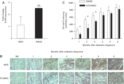
Fig. 3.
TLR4 is increased and lipid accumulates in the cardiac muscle of diabetic mice. A: quantitative PCR was carried out in age-matched nondiabetic (ND) and recent-onset (within 3 wk) DNOD mice (16–18 wk of age) for the expression of TLR4 in cardiomyocytes. **P < 0.05. B: lipid accumulation in the ventricular muscle of diabetic mice over time after the diagnosis of diabetes, as shown by oil red O staining (brown). C: lipid staining of heart muscle shown in the frozen tissue sections in A was quantified, and the results are shown in arbitrary units. *P < 0.05, DNOD mice vs. DTLR4KO mice in the first 3 mo after the diagnosis of diabetes. Diabetic mice were 12–14 wk of age at the time of diagnosis, and control ND mice were age matched.

