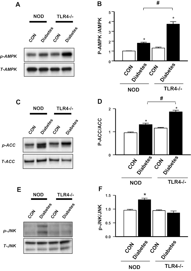
Fig. 5.
Expression of AMP-activated protein kinase (AMPK), acetyl-CoA carboxylase (ACC), and JNK in cardiomyocytes of DNOD mice compared with DTLR4KO mice. A: Western blot analysis for p-AMPK and total (T-)AMPK in ventricular muscle samples taken at 2–3 mo after the diagnosis of diabetes showing control nondiabetic mice compared with diabetic mice for both WT and TLR4KO strains. B: densitometry readings comparing p-AMPK with total AMPK from the samples shown in A. n = 3 mice/group. *P < 0.05, control mice vs. diabetic mice; #P < 0.05, DNOD mice vs. DTLR4KO mice. C: Western blot analysis for p-ACC and total ACC in ventricular muscle samples taken at 2–3 mo after the diagnosis of diabetes showing control nondiabetic mice compared with diabetic mice for both WT and TLR4KO strains. D: densitometry readings comparing p-ACC with total ACC from the samples shown in C. n = 3 mice/group. *P < 0.05, control mice vs. diabetic mice; #P < 0.05, DNOD mice vs. DTLR4KO mice. E: Western blot analysis for p-JNK and total JNK in ventricular muscle samples taken at 2–3 mo after the diagnosis of diabetes showing control nondiabetic mice compared with diabetic mice for both WT and TLR4KO strains. F: densitometry readings comparing p-JNK with total JNK from the samples shown in E. n = 3 mice/group. *P < 0.05, DNOD mice vs. other groups. Diabetic mice were 12–14 wk of age at the time of diagnosis of diabetes, and control mice were age matched.

