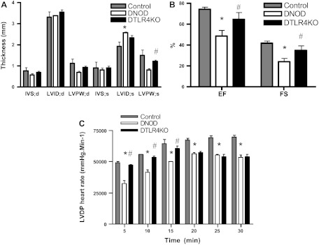
Fig. 8.
Cardiac functional abnormalities were attenuated in DTLR4KO mice. A and B: echocardiograms were performed on nondiabetic control (n = 3), DNOD (n = 6), and DTLR4KO (n = 7) mice 2–3 mo after the diagnosis of diabetes. A: interventricular septum at diastole (IVS:d), interventricular septum at systole (IVS:s), left ventricular (LV) posterior wall at diastole (LVPW:d), LV posterior wall at systole (LVPW:s), LV internal dimension at diastole (LVID:d), and LV internal dimension at systole (LVID:s). B: ejection fraction (EF; in %) and fractional shortening (FS; in %). C: hearts were isolated and perfused for 30 min, and LV developed pressure (LVDP) multiplied by heart rate (mmHg·beats·min−1) was measured. *P < 0.05, DNOD mice vs. control mice; #P < 0.05, DTLR4KO mice vs. DNOD mice. Diabetic mice were 12–14 wk of age at the time of diagnosis of diabetes, and control mice were age matched at the time of measurement.

