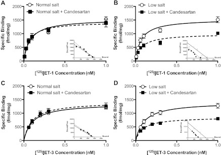
Fig. 5.
Saturation binding curves for [125I]-ET-1 (A and B) or [125I]-ET-3 (C and D) in plasma membrane preparations of renal inner medullary tissue from rats on a normal (A and C)- or low (B and D)-salt diet with or without candesartan. Insets: Scatchard analysis of [125I]-ET-1 and [125I]-ET-3; n = 3–4 rats/group.

