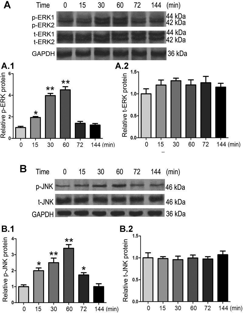
Fig. 4.
Effect of TGF-β1 on the phosphorylation of ERK1/2 and JNK MAPK in HK-2 cells. Cells were exposed to TGF-β1 (10 ng/ml) for varying periods of time, cellular extracts were subjected to SDS-PAGE, and their protein blots were probed with various antibodies. A and B: TGF-β1 treatment increased ERK1/2 (A) and JNK (B) phosphorylation (p-ERK1/2 and p-JNK) in a dose-dependent manner with a peak effect around 60 min after exposure to the cytokine. No changes were observed in total (t-)ERK1/2 and JNK. The bar graphs (1 and 2) represent densities of the corresponding bands detected by immunoblot procedures relative to GAPDH. Values are expressed as means ± SE; n = 5. *P < 0.05 and **P < 0.01 compared with the control group.

