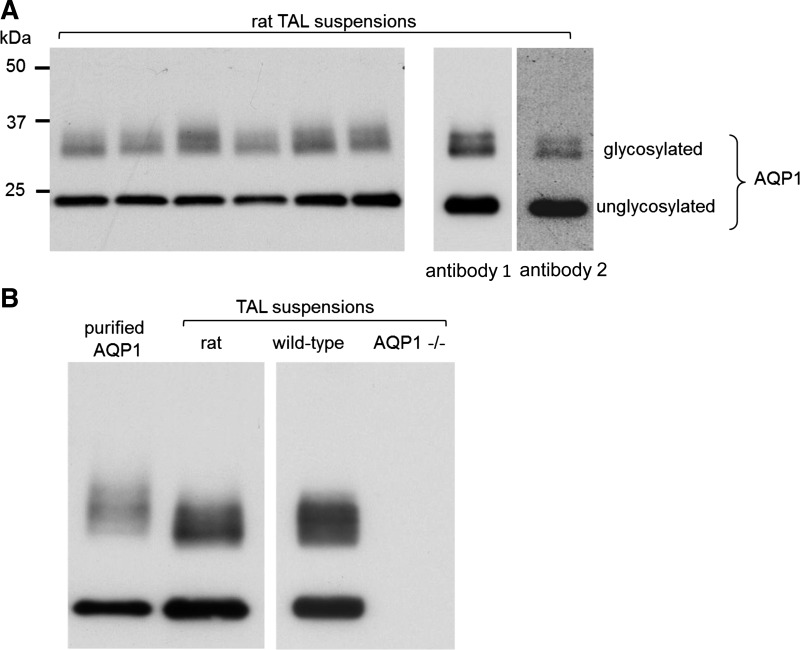
Fig. 1.
Detection of aquaporin (AQP)1 in rat thick ascending limb (TAL) suspensions using Western blots. A: 10-μg suspensions from different animals were loaded into each lane (n = 6); right: identical sized bands were obtained using 2 distinct anti-AQP1 antibodies. Here, the membrane was cut and each piece was used to test the different antibodies (brightness and contrast were enhanced in this particular blot to make the bands visible on the picture). B: purified AQP1 as a positive control; right: TALs from Aqp1 −/− mice as a negative control. WT, wild-type. Representative blots from different gels are separated by the white space between them.

