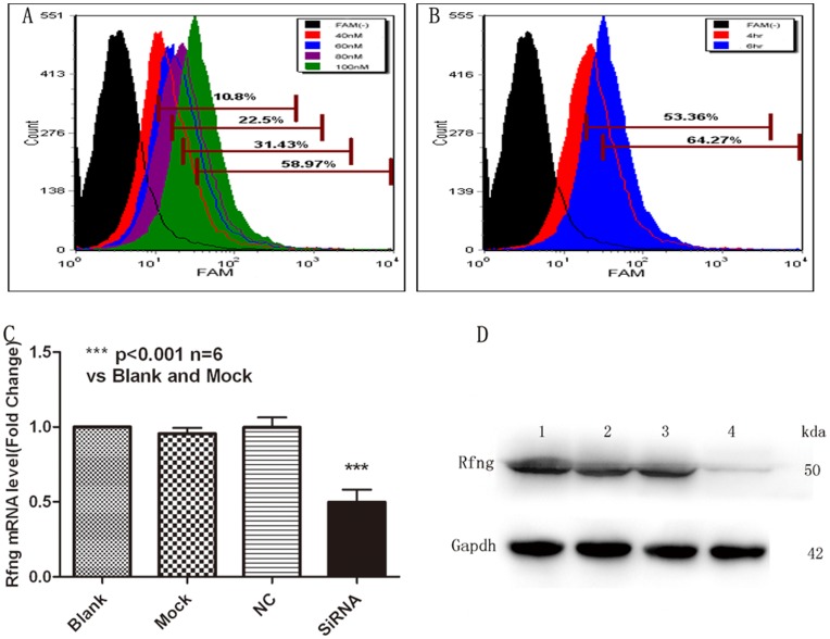
Figure 4. Asthmatic naïve CD4+T cells are efficiently transfected with SiRNA.
CD4+T (5×10⁶) cells were transfected by Amaxa Nucleofection, protocol U-14, with 40 nM, 60 nM, 80 nM, 100 nM FAM-tagged SiRNA. The transfection conditions optimized by flow cytometry analysis. Q-PCR and Western blot were performed to determined the mRNA levels and protein levels after SiRNA-Rfng transfection. A, The optimal final concentration of SiRNA was 100nM (the MOI is 58.97%). B, The optimal time of transfection is more than 6 hr (64.27%). C, Real-time PCR analysis of Rfng levels in transfected naïve CD4+T cells. Blank-treated results were taken as 1. Results are from three independent experiments. The data for each group are expressed as means±SEM. *** p<0.05, significant differences between siRNA-Rfng and blank-treated, mock-treated or SiRNA-scrambled CD4+T cells. D, Rfng protein levels in transfected CD4+T cells. CD4+T cells were unmanipulated (blank, lane 1), transfected with reagent alone (mock, lane 2), siRNA scrambled (lane 3), or siRNA-Rfng (Lane 4). Transfected CD4+T cells were lysates and collected to assess the expression of Rfng by Western blot. Gapdh was used as a loading control. Representative of one of three similar experiments.

