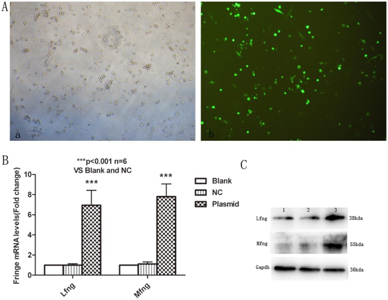
Figure 5. Overexpression of Lfng and Mfng in naïve CD4+T cells. A.
, Asthmatic naïve CD4+T cells were transfected with pEGFP-N1 plasimd using Amaxa Nucleofection System. 5×10⁶ cells were resuspended in 100 µl of the appropriate Amaxa solution and transfected with 5 µg pEGFP-N1 plasimd. 6–8 hrs’ later, GFP expression was detected under fluorescence microscope to optimize the transfection conditions. The optimal transfection efficiency was approximately 80% (a, bright field; b, fluorescence field). Original magnification was×100. B, Real-time PCR analysis of Lfng and Mfng levels in transfected CD4+T cells. Blank-treated results were taken as 1. Results are from three independent experiments. The data for each group are expressed as means±SEM. *** p<0.001, significant differences between plasmid overexpression group and blank group, or NC (pEGFP-N1) group CD4+T cells. C, Lfng and Mfng protein levels in transfected CD4+T cells. CD4+T cells were unmanipulated (Blank, lane 1), transfected with pEGFP-N1 plasmid (NC, lane 2), or transfected with Lfng plasmid or Mfng plasmid (Lane 4). Representative of one of three similar experiments.

