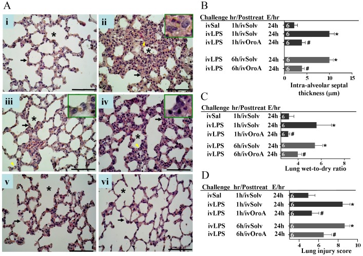
Figure 2. Inhibition of LPS-induced lung inflammation by OroA post-treatment.
Representative sections of the rat lung tissues were stained with hematoxylin and eosin. In control section (panel Ai), normal alveoli (asterisk) and alveolar septa (arrow) with few neutrophils were shown. 24 hrs after LPS treatment (10 mg/kg, iv), thickened septa were observed in panel Aii, and inset is the enlarged area (indicated by arrowhead) of activated alveolar macrophages. Oro-A (15 mg/kg, iv) given 1 hr (panels Aiii and B) and 6 hrs (panels Aiv and B) after LPS treatment significantly reversed thickened septa when examined 24 hrs after LPS challenge. This concentration of Oro-A given 1 hr (panel Av) and 6 hrs (panel Avi) after saline (Sal) treatment did not show any effect when examined 24 hrs after LPS challenge. Panel C indicates that the enhanced lung wet-to-dry weight (W/D) ratio following LPS treatment (10 mg/kg, iv) was reversed by OroA treatment (15 mg/kg, iv) administered 1 hr or 6 hrs after LPS challenge. In panel D, LPS (10 mg/kg, iv) significantly increased lung injury score comparing to that of Sal (normal saline) control when examined 24 hrs (E/24h) after LPS challenge. The increase was reversed significantly by OroA (15 mg/kg, iv) administered 1 hr or 6 hrs after LPS challenge. Solv, normal saline plus Tween 80 at 9∶1 ratio; hr/Posttreat (post-treatment hour after LPS treatment); E/hr (examination hour after LPS challenge). Data are means±SEM. *P<0.05 indicates significant difference from the normal control (ivSal-ivSolv) group. #P<0.05 indicates significantly different from the respective (1 hr or 6 hrs) LPS alone (ivLPS-ivSolv) group. The number in each column represents the number of rats used. Scale bar, 50 µm.

