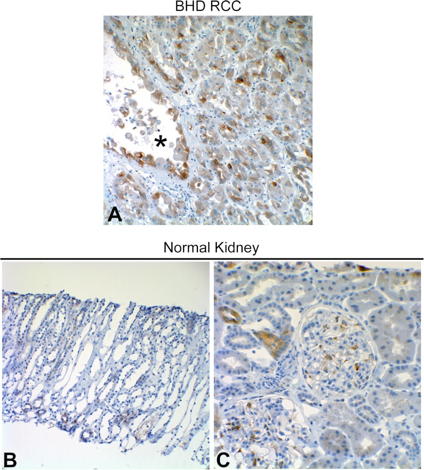
Fig. 11.
A: immunohistochemical analysis of BiP expression in renal cell carcinomas (RCC) and RCC cyst (lumen marked with *) tissue from a patient with Birt-Hogg-Dubé syndrome. Lower magnification (B) and higher magnification (C) immunohistochemical images of normal renal tissue (obtained via core needle biopsy) stained for BiP.

