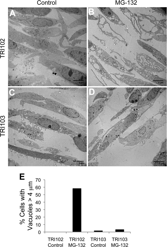
Fig. 4.
Electron micrographs of TRI102 (A and B) and TRI103 (C and D) cells untreated or treated with MG-132 (500 nM, 8 h). Note the pronounced vacuolization in MG-132 treated TRI102 cells (B) compared with other groups. E: graph depicting the percentage of cells containing vacuoles >4 μm in longest diameter. A minimum of 50 cells were counted for each group.

