Fig. 11.
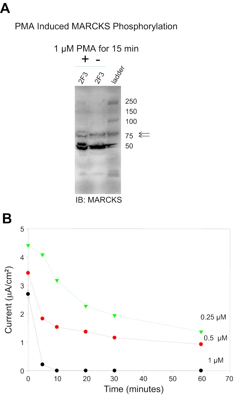
Measurements of transepithelial current in Xenopus 2F3 cells treated with PMA. A: Western blot analysis showing PMA-induced phosphorylation of MARCKS after 15 min using a polyclonal antibody specific for MARCKS (arrows) and MARCKS-related protein (MRP; bottom bands). The top band of the doublet represents the phosphorylated form of MARCKS. B: transepithelial resistance and voltage were measured after application of PMA on the apical side of Xenopus 2F3 cells, and transepithelial current was calculated. PMA treatment resulted in a dose- and time-dependent decrease in transepithelial current.

