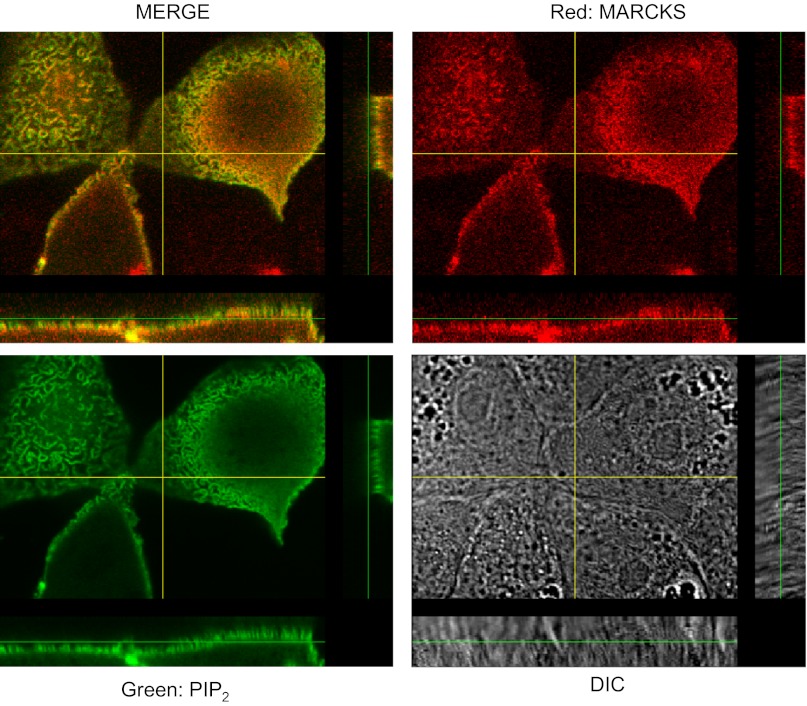
Fig. 7.
Localization of MARCKS and PIP2. 2F3 cells were transfected with the PIP2 reporter GFP-PLC-δ1 PH domain, fixed in paraformaldehyde, and stained with primary antibodies against MARCKS protein (host: mouse). Following treatment with a fluorescent secondary antibody, cells were examined using confocal microscopy using an Olympus Fluoview 1000 confocal microscope. The 4 panels show an X-Y optical slice near the apical membrane showing PIP2 (green), MARCKS (red), the merged image, and a differential interference white light image. Also, z-axis images are shown adjacent to the X-Y images. The overlap of red and green is best seen in the z-axis images.

