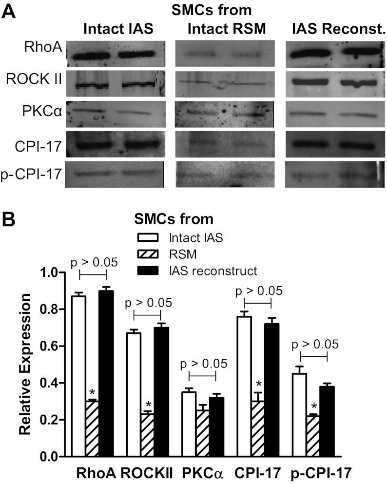
Fig. 6.
A: Western blots (WB) of RhoA, ROCKII, PKCα, protein kinase C-potentiated inhibitor or inhibitory phosphoprotein for myosin phosphatase (CPI-17), and phospho (p)-CPI-17 in the smooth muscle cells (SMCs) isolated from intact IAS, intact rectal smooth muscle (RSM), and the IAS reconstructs. B: respective quantitative data. Data show significantly higher levels of above signal transduction protein with the exception of PKCα, in the cells from intact IAS, and constructs compared with those from intact RSM (*P < 0.05; n = 5).

