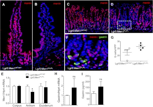
Fig. 11.
Epithelium-specific Men1 deletion does not lead to complete menin deletion in G cells. A and B: representative images of menin (red) immunofluorescent staining in sections of duodenum from Lgr5:Men1WT/WT and Lgr5:Men1FL/FL mice 5 mo after tamoxifen administration. Nuclei were stained with DAPI (blue). Original magnification: ×200. C and D: coimmunostaining of paraffin sections for menin (red) and gastrin (green) confirms colocalization of these proteins to antral G cells of Lgr5:Men1WT/WT and Lgr5:Men1FL/FL mice. Nuclei were stained with DAPI (blue). Original magnification: ×200. E: RT-qPCR measurement of Men1 mRNA normalized to Hprt mRNA levels in the corpus, antrum, and duodenum of Lgr5:Men1FL/FL and Lgr5:Men1WT/WT mice. Values are means ± SE; n = 3. *P < 0.05. F: higher-power magnification (×600) of D. G: morphometric analysis of G cell number in Lgr5:Men1WT/WT and Lgr5:Men1FL/FL mice 5 mo after tamoxifen administration. Values are numbers of gastrin-immunostained cells per HPF (×400 magnification). H: RT-qPCR measurement of gastrin mRNA normalized to Hprt mRNA levels in the stomach of Lgr5:Men1FL/FL and Lgr5:Men1WT/WT mice. Values are means ± SE; n = 3. I: enzyme immunoassay measurement of gastrin concentration in circulating plasma of mice fasted for 16 h. Values are means ± SE; n = 3.

