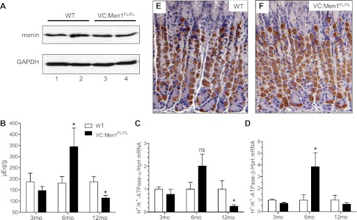
Fig. 9.
Menin expression is not altered in the corpus of VC:Men1FL/FL mice. A: Western blot analysis of menin protein in the corpus of 2 WT (lanes 1 and 2) and 2 VC:Men1FL/FL (lanes 3 and 4) mice at 12 mo of age. B: basal gastric acid levels in 3-, 6-, and 12-mo-old fasted WT and VC:Men1FL/FL mice. Values are means ± SE; n = 4–13. *P < 0.05. C and D: relative H+-K+-ATPase-α and -β mRNA expression measured by RT-qPCR from the gastric mucosa of WT and VC:Men1FL/FL mice and normalized to Hprt mRNA. Values are means ± SE; n = 3–4. *P < 0.05. E and F: paraffin sections immunostained with H+-K+-ATPase-α antibodies showing parietal cells in the corpus of WT and VC:Men1FL/FL mice.

