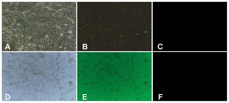
Figure 1. Morphological identification of cultured marginal cells and 3T3 cells.
A, Under phase contrast microscopy, marginal cells were arranged like paving stones, and displaying a polygonal shape and large nuclei (×100). B, Under fluorescence microscopy, numerous star-like green pots were observed within the cytoplasm (×100). C, Negative control of marginal cells (background fluorescence). D, Under phase contrast microscopy, 3T3 cells were arranged spindle -shaped flat structure with cell protrusions and large nuclei(×40). E, Under fluorescence microscopy, no positive green staining was observed within the cytoplasm (×40). F, Negative control of 3T3 cells (background fluorescence).

