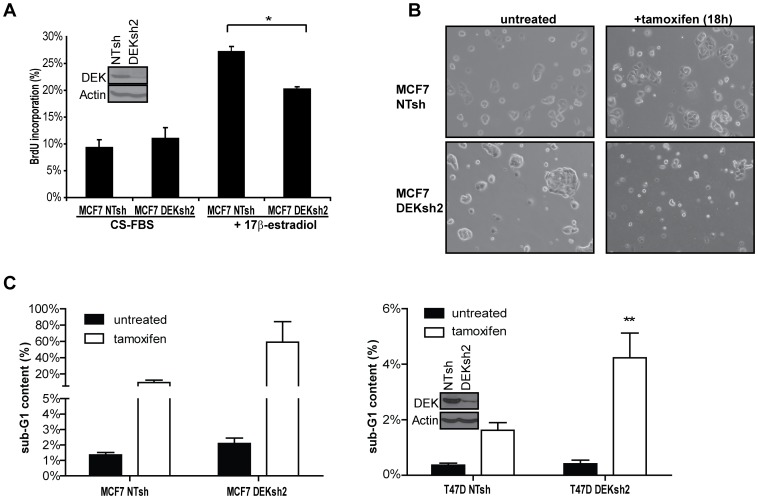
Figure 3. DEK is necessary for 17β-estradiol stimulated cell proliferation and modulates sensitivity to tamoxifen.
(A) DEK expression is required for 17β-estradiol stimulated cellular proliferation. Hormone starved MCF7 cells transduced with non-targeting shRNA (NTsh) or DEK shRNA (DEKsh2) were untreated (CS-FBS) or exposed to 10 nM 17β-estradiol, then cultured in BrdU. The percentage of BrdU positive cells was determined by flow cytometry. Asterisk (*) denotes p<0.05 using Student’s t-test. (B and C) DEK depletion by shRNA (DEKsh2) works synergistically with tamoxifen to induce apoptosis in breast cancer cell lines. (B) Bright field images (100× magnification) of MCF7 cells expressing either NTsh or DEKsh2 were cultured in low serum media and either untreated or treated with tamoxifen for 18 hours. (C) DEK depletion by shRNA (DEKsh2) enhances the cytotoxic effect of tamoxifen. DEK proficient and deficient MCF7 (left) and T47D (right) cells were grown in low serum media then treated with 3 µg/ml tamoxifen for 22 hours. Cells were labeled with 7AAD then analyzed for sub-G1 content by flow cytometry as a measure of apoptosis. Results shown are the average of triplicate experiments. Two asterisks (**) indicate p<0.01 as determined using a 2-way ANOVA test for significance. For MCF7 cells, p = 0.08. (A and B insets) DEK shRNA knockdown is shown by western blot analysis for normally cultured cells that were transduced with lentivirus carrying either non-targeting shRNA (NTsh) or DEK specific shRNA (DEKsh2).

