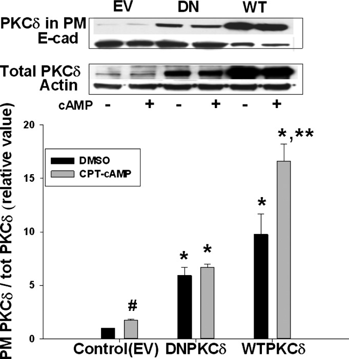
Fig. 5.
DN- as well as WT-PKCδ increased PM PKCδ. HuH-NTCP cells were transfected with EV, DN-PKCδ, or WT-PKCδ, followed by treatment with or without 100 μM CPT-cAMP for 15 min. A biotinylation method was used to determine PM PKCδ. Representative immunoblots of PM PKCδ (top) and total PKCδ (middle) and the densitometric analysis (bottom). Amount of PKCδ localization in the PM was expressed as a ratio of PM PKCδ to total PKCδ. Relative values of PKCδ in the PM are expressed as means ± SE (n = 3). Data were analyzed by 1-way ANOVA. *Significantly different (P < 0.05) from control values in the absence of cAMP; **significantly different (P < 0.05) from values in the absence of cAMP in cells transfected with WT-PKCδ. Control values in the absence and presence of cAMP are #significantly different (P < 0.05) by paired t-test.

