Fig. 1.
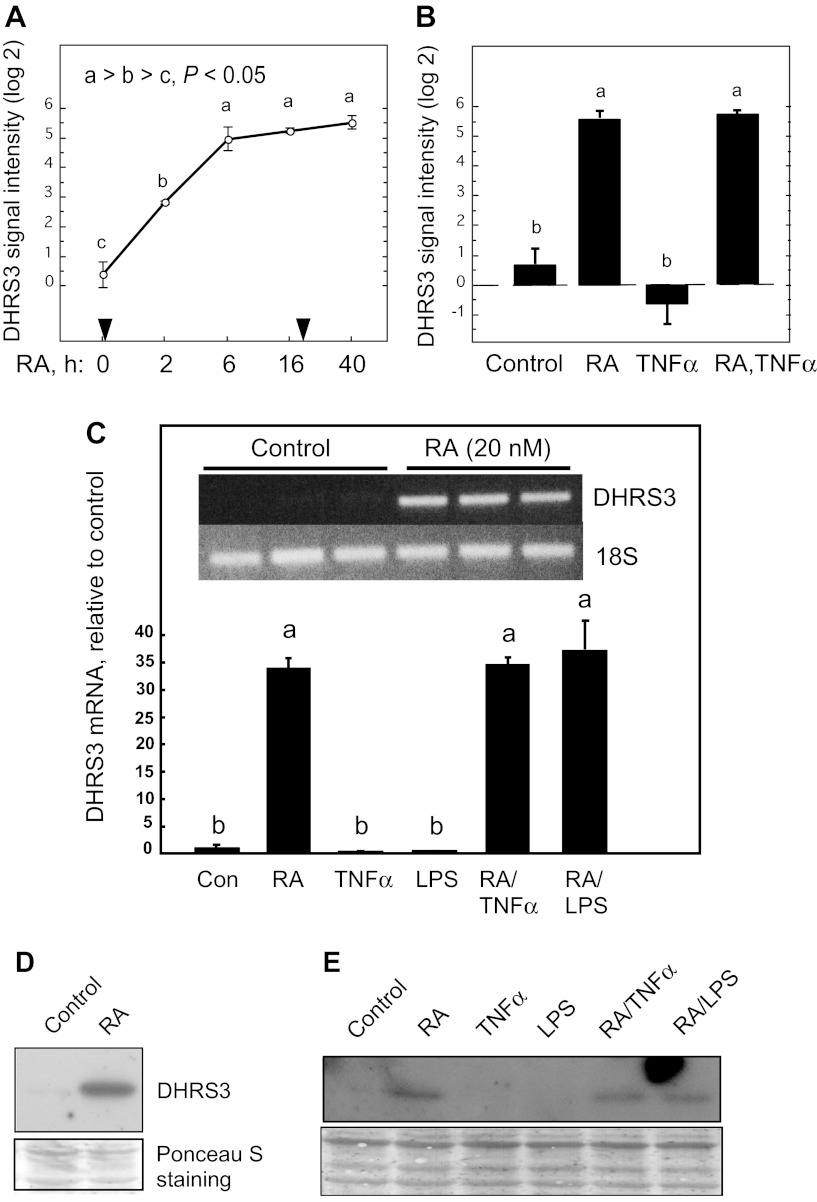
Expression and regulation of DHRS3 mRNA in THP-1 cells treated with retinoic acid (RA), TNFα, or LPS, and combinations of these agents. A: DHRS3 mRNA in THP-1 cells treated with 20 nM RA at time 0; an equal amount of RA was added after 24 h of incubation. B: DHRS3 mRNA after 40 h of treatment with RA, TNFα (5 ng/ml, added 16 h after time 0), and RA (added at time 0) followed by TNFα (added at 16 h for a total of 40 h). Results in A and B represent 2–6 replicates, determined by microarray analysis. For 2 different target sequences of DHRS3 mRNA, log2 difference (maximum − minimum) was 5.16 (A) and 4.16 (not shown), respectively. C: relative intensity of DHRS3 mRNA in THP-1 cells treated with vehicle or RA (20 nM), each in triplicate, for 6 h and then harvested for RNA analysis by semiquantitative RT-PCR. Inset: ethidium bromide-stained agarose gel. Treatments were as follows: vehicle only (Con), RA (20 nM), TNFα (5 ng/ml), LPS (100 ng/ml), RA + TNFα, and RA + LPS (n ≥ 3/group). D: DHRS3 protein detected by immunoblot on pooled cell extracts using human DHRS3 monoclonal antibody. THP-1 cells were cultured with vehicle only (control) and with RA (20 nM, 6 h). Ponceau S staining was used as a loading control. E: DHRS3 protein detected by immunoblot using human DHRS3 monoclonal antibody. Cell extracts were prepared from THP-1 cells treated as described in C. Ponceau S staining was used as a loading control. Differences were determined by 1-way ANOVA: different letters (a, b, c) indicate significant difference: P < 0.05.

