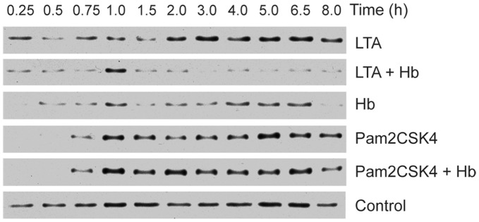
Figure 5. Degradation of IκBα during macrophage activation.
HeNC2 cells were stimulated with LTA (1 µg/ml), LTA plus Hb (1 µg/ml and 50 µg/ml, respectively), Hb (50 µg/ml), Pam2CSK4 (100 ng/ml), Pam2CSK4 plus Hb (100 ng/ml and 50 µg/ml, respectively) or maintained in medium for the indicated times. Cell extracts were electrophoresed on SDS-PAGE gels, transferred to nitrocellulose and incubated with anti-IκBα. Similar results were obtained from each of two independent experiments.

