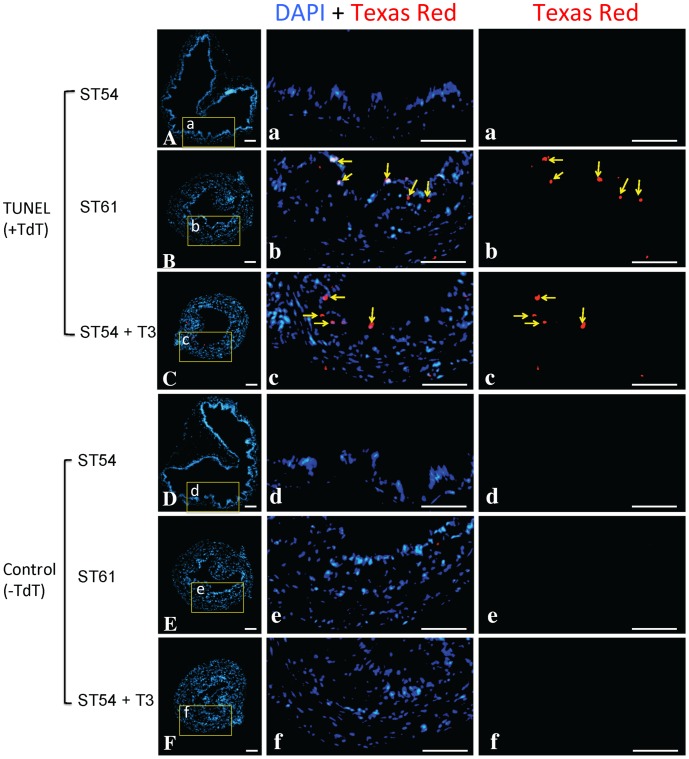
Figure 6. Intestinal epithelial apoptosis occurs during natural and T3-induced metamorphosis in X. tropicalis.
The intestine from tadpoles at premetamorphic stage 54 (ST54), the climax of metamorphosis (ST61), or ST54 but treated with 5 nM T3 for 3 days (ST54+ T3), were analyzed by TUNEL assay (A–C) or TUNEL without the enzyme TdT as a the negative control (D–F). The biotin-dUTP labeled apoptotic cells were visualized with Texas-Red labeled Streptavidin and the nuclei were stained with DAPI. The boxed areas were shown at a higher magnification (a-f) with the apoptotic cells indicated with yellow arrows in either merged (DAPI+Texas Red) and single channel of red fluorescence (Texas-Red) images. Scale bar: 50 µm.

