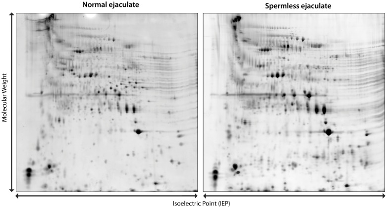
Figure 3. Two-dimensional gel electrophoresis of male ejaculates.
Protein gels of a normal male ejaculate (left) and a spermless male ejaculate (right). Spermatophore samples were ground and centrifuged to remove spermatophore debris and sperm. Proteins were separated based on their isoelectric point in the first dimension and molecular weight in the second.

