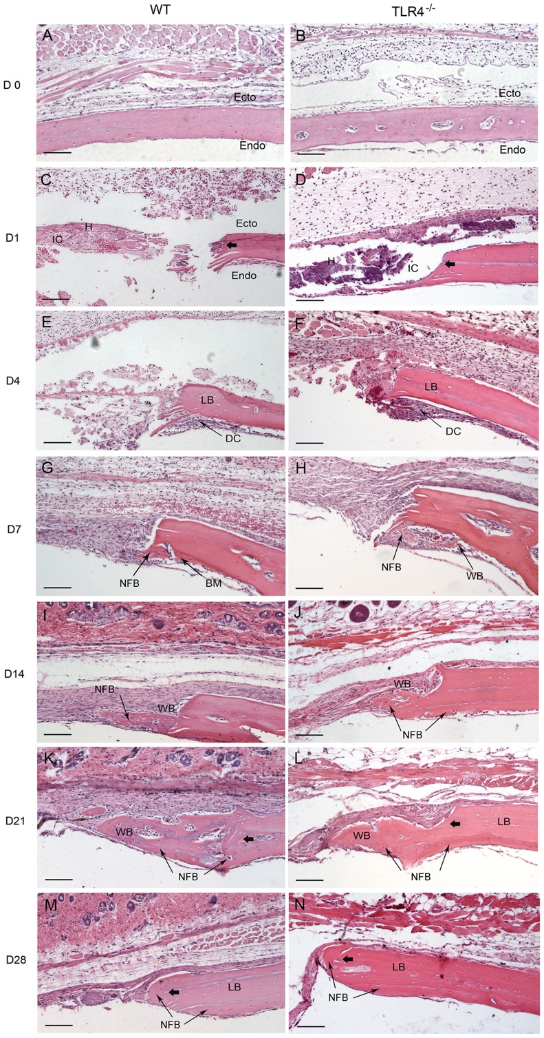
Figure 1. Histophotomicrographs for H&E stained tissues at the defect margins at postoperative time points.
WT and TLR4−/− mice showed similar histological staining patterns on days 0, 1 and 4, while larger areas of newly-formed bone were seen in TLR4−/− mice than in WT mice on day 7. Newly-formed cellularized bone matrix was observed on the endocortical (dural) side of the calvarial bone lateral to the defect perimeter in both groups since day 7. Active bone formation was suggested by the presence of large regions of woven bone matrix at the defect margin in both groups on days 14 and 21. Defects in WT and TLR4−/− mice were histologically similar since day 21. (scale bar: 100 µm; bolded arrows: defect margin; endo: endocortical surface of calvarial bone; ecto: ectocortical surface of calvarial bone; H: hematoma; IC: infiltrating cells; LB: lamellar bone; WB: woven bone; NFB: newly-formed bone; DC: dural cells).

