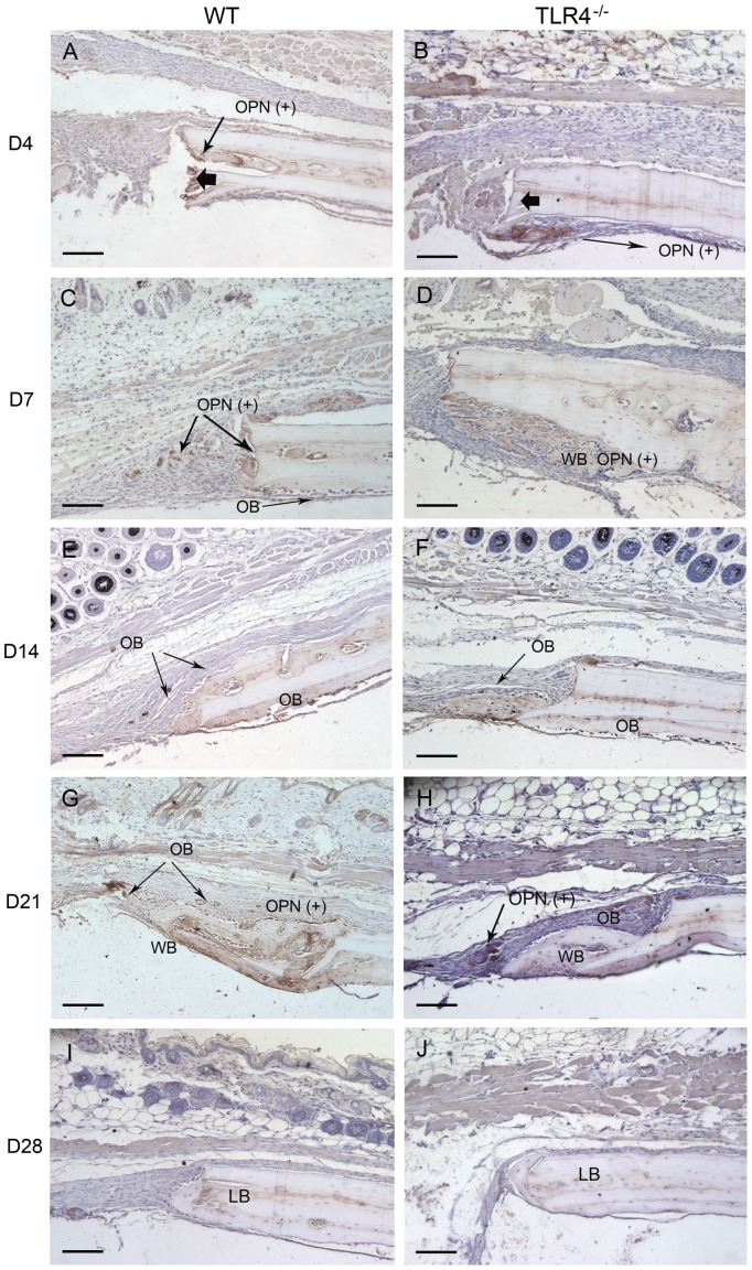
Figure 3. Histophotomicrographs of pentachrome stained tissues in WT and TLR4−/− mice.
Similar stains were observed between WT and TLR4−/− mice on day 0 and day 1. An increased amount of newly-formed bone was observed in TLR4−/− mice at day 7, suggesting accelerated healing compared to WT. Lamellar bone (*), which stains positive for acid fuchsin (red) was observed in both groups on day 28, suggesting maturation and remodeling of the newly formed bone matrix. (scale bar: 100 µm; bolded black arrows: defect margin; LB: lamellar bone; NFB: newly-formed bone).

