Figure 3.
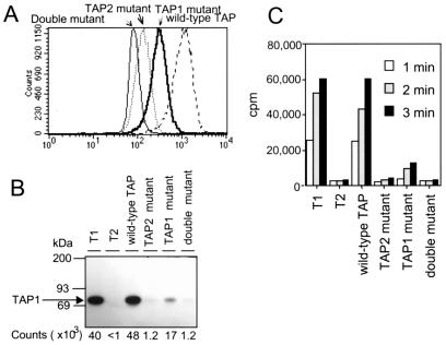
Reconstitution of T2 cells with wild-type and mutant TAP polypeptides. (A) MHC class I expression in wild-type and mutant TAP-transfected T2 cells. Wild-type and mutant TAP-transfected T2 cells were analyzed for MHC class I expression (HLA-B alleles) by flow cytometry, using the monoclonal antibody 4E. (B and C) Peptide binding and translocation require a catalytically active NBD on TAP2. (B) Photolabeling of TAP from wild-type and mutant TAP-transfected T2 cells with the [125I]KB11-HSAB peptide was performed as described in Materials and Methods. After solubilization in 1% digitonin, labeled TAP molecules were immunoprecipitated with the anti-TAP1 mAb. 148.3 and analyzed by SDS/PAGE and autoradiography, and bands corresponding to 125I labeled TAP1 were quantitated by PhosphorImager analysis. (C) SLO permeabilized cells from wild-type and mutant T2 TAP transfectants were incubated with the iodinated reporter peptide RRYQNSTEL at 37°C for the indicated time periods, and the reaction stopped by lysis with cold 3% Triton X-100, as described in Materials and Methods. Translocation into the ER was assessed by binding of the glycosylated reporter peptide to Con A-Sepharose beads and counting on a γ-counter.

