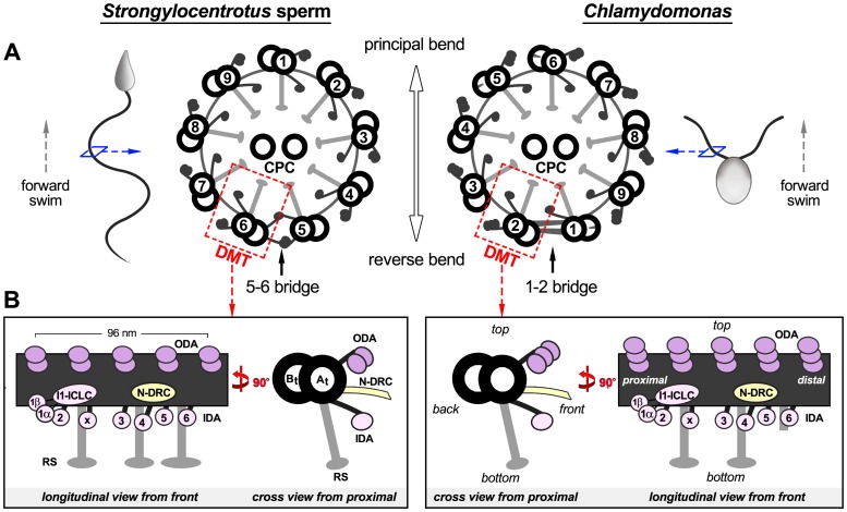
Figure 1. Schematic models of the axoneme organization in the sea urchin Strongylocentrotus and the green alga Chlamydomonas.
(A) Two numbering systems for designating the DMTs in 9+2 cilia and flagella are currently in use (left [7], right [15]). The flagella from both organisms have a highly conserved cylindrical arrangement of nine DMTs (red boxes). Each DMT is built from many copies of a 96-nm long unit that repeats along the DMT length. The axonemes are shown in cross-sectional views from the flagellar base (proximal) towards the tip (distal). The locations of the previously described 5–6 bridge (left) and proximal 1–2 bridge (right) are indicated. (B) For both organisms, schematic representations of a 96-nm repeat are shown in longitudinal and cross-sectional views; orientations of the 96-nm repeat are maintained in all following figures unless stated otherwise. Other labels: A-tubule (At), B-tubule (Bt), inner dynein arms (IDA 1α, 1β, 2–6 and x; rose) [43], intermediate chain/light chain complex (ICLC), nexin-dynein regulatory complex (N-DRC, yellow) [37], outer dynein arm (ODA, purple), radial spoke (RS, gray) [13], [30].

