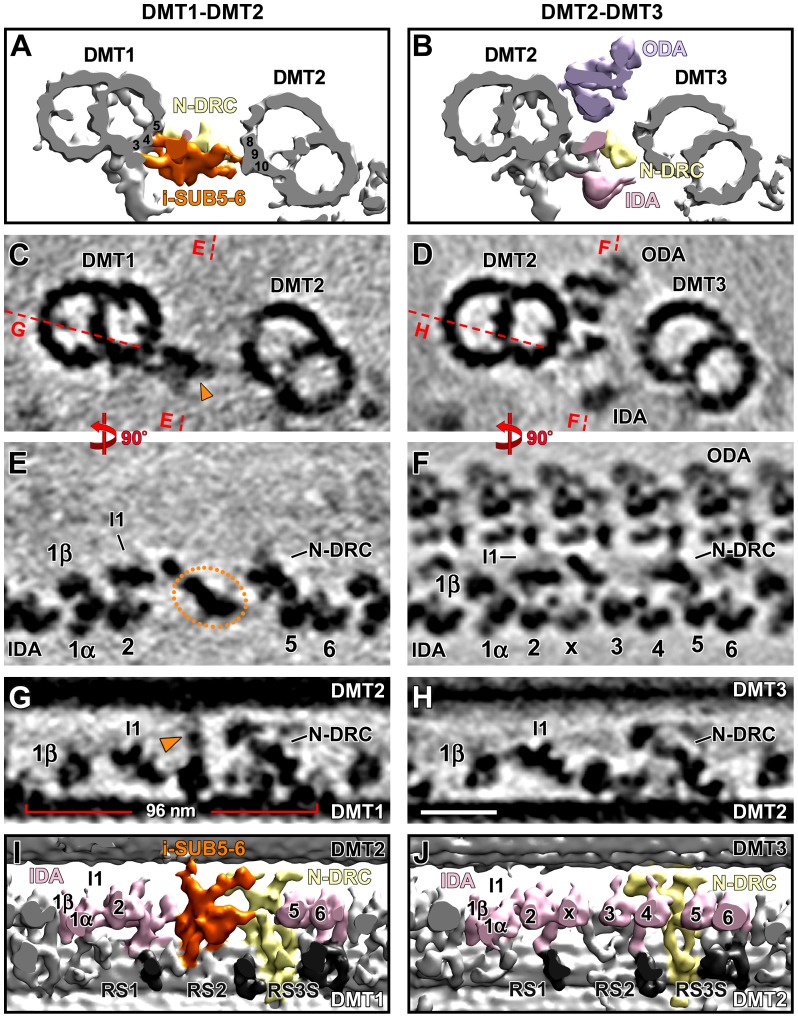
Figure 3. Distinct structural features in the distal three quarters of Chlamydomonas DMT1.
Isosurface renderings (A, B, I, and J) and tomographic slices (C–H) show i-SUB5-6 and the missing IDAs on Chlamydomonas DMT1 (left column) in cross-sectional (A–D) and longitudinal views from the front (E and F) and the bottom (G–J). The structure of DMT1 is unique and different from DMTs 2–9, which have a similar structure; DMT2 is shown for comparison in the right column (see also Figure S1 and Movie S3). (A–D) Cross-sectional views of the i-SUB5-6 complex (orange), which is present on DMT1 only and links DMT1 to DMT2. The ODAs are missing from DMT1 but are present on all other DMTs. The red dashed lines in (C and D) indicate the locations of the tomographic slices shown in (E–H). Note that the beak structure in the B-tubule of DMT1 in (A and C) has previously been reported to be present in the proximal half of the flagellum [15], and is therefore still visible in this average of the distal three quarters of the axoneme. (E and F) Several IDAs (IA3, 4, and IAX) are absent on DMT1 (E) but present on DMT2 (F). The orange dotted circle in (E) outlines the i-SUB5-6 complex that is present on DMT1, but absent from DMT2 (F). (G–J) Details of the i-SUB5-6 structure (orange) demonstrate that it has a periodicity of 96 nm and makes clear connections with both I1 dynein and N-DRC while linking DMT1 to DMT2. Other labels: A-tubule (At), B-tubule (Bt), inner dynein arms (IDA 1α, 1β, 2–6 and x; rose), nexin-dynein regulatory complex (N-DRC, yellow), outer dynein arm (ODA, purple), radial spoke (RS, dark-gray). The Chlamydomonas DMT numbers are according to Hoops and Witman [15]. The protofilaments are numbered after Linck and Stephens [53]. Scale bar (H): 25 nm.

