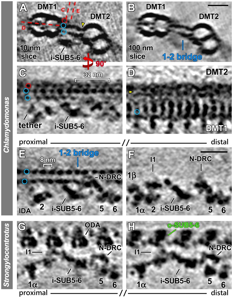
Figure 4. Distinct structural features in specific regions of Chlamydomonas DMT1 and Strongylocentrotus DMT5.
(A–E) The 1–2 bridge in the proximal quarter of Chlamydomonas DMT1. Tomographic slices show the proximal 1–2 bridge between DMTs 1 and 2 in Chlamydomonas pWT in cross-sectional (A and B) and longitudinal views from the front (C and E) and the bottom (D). Red dashed lines in (A) indicate the locations of tomographic slices shown in (C–E). The 1–2 bridge consists of two parts that link DMTs 1 and 2; each part consists of a row of periodic densities (blue circles in A, and C–E). The connection of the top row to DMT2 is highlighted (yellow arrowheads in A and D). The red circles in (A and C) indicate an additional row of periodic but shorter densities that do not reach DMT2. Note that the I1 dynein is absent from the proximal quarter of DMT1 and only a small neighboring density, the I1 tether [34], is visible (C and E). (F) A tomographic slice from the distal part of DMT1 is shown for direct comparison with the proximal slice in (E). Note that the distal region of Chlamydomonas DMT1 lacks the 1–2 bridge, while the I1 dynein is present. (G and H) Tomographic slices in front view show a comparison between the proximal 1/8th (G) and distal 7/8th (H) of the Strongylocentrotus DMT5. While in the very proximal region regular ODAs are present, they are substituted by o-SUB5-6 structures in the distal 7/8th of the flagellum. Note that along the entire length of DMT5 i-SUB5-6 is consistently present, while several IDAs (IA 2–4, IAX) are lacking. Scale bars are 25 nm (scale bar in B valid for A and B; scale bar in F valid for C–H).

