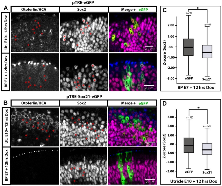
Figure 5. Short induction of Sox21 reduces Sox2 expression in organotypic cultures of E10 utricle and E7 basilar papilla.
(B) Representative images of Sox2 immunostaining in pTRE-Sox21-eGFP samples after 12 hours of Dox treatment. Induced cells are marked with asterisks, and tend to exhibit reduced levels of Sox2 expression compared to neighbouring untransfected cells (C). Box plot of Z-score values [Z = (x-mean)/stdev] for levels of Sox2 expression in supporting/progenitor cells of the basilar papilla (B; 3 samples) or of the utricle (C; 5 samples), expressing either eGFP only or Sox21-eGFP. Outliers, minimum, first quartile, median, third quartile and maximum are displayed, n = numbers of transfected cells. Sox2 expression levels were significantly lower in Sox21-eGFP expressing cells than in eGFP expressing cells in both the auditory (Mann-Whitney U = 14873; p = 0.00) and vestibular (Mann-Whitney U = 32335; p = 0.00) epithelia.

