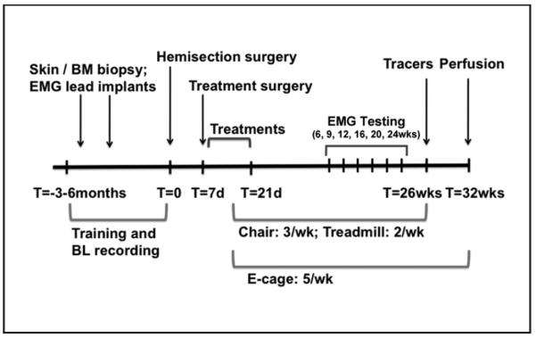
Figure 1.
Timeline: T = 0 indicates the time of spinal cord hemisection. In a period of 3 to 6 months before spinal cord hemisection, animals underwent 2 preparatory surgeries: a skin or bone marrow (BM) biopsy and implantation of EMG leads. In addition, animals were trained to perform chair, treadmill, and open-field tasks. Training and collection of baseline (BL) data on these tasks occurred up until the week of spinal cord hemisection. Then, 7 days after spinal cord hemisection, animals underwent a treatment surgery followed by 14 days of treatment delivery in the hospital and home cage. Behavioral testing in the chair (3 times weekly), treadmill (twice weekly), and open field (5 times weekly) resumed once animals were ready (during week 2 after spinal cord hemisection) and continued until week 25 (chair and treadmill) and week 32 (open field). EMG and 3D video recording sessions occurred before the lesion and at weeks 6, 9, 12, 16, 20, and 24. Animals underwent surgery 6 weeks prior to perfusion for delivery of tracers into the motor cortex, brainstem, and spinal cord.

