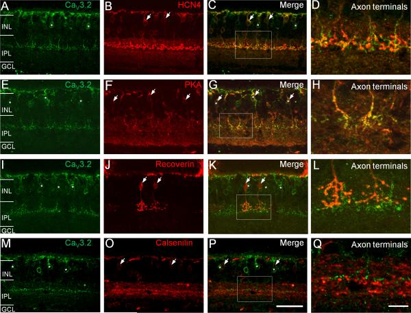
Figure 3. Double-labeling analysis of Cav3.2-positive bipolar cells in wild-type mouse retinas.
(A–D) Cav3.2-positive bipolar cells (A, green, asterisks) do not show labeling by an antibody against HCN4 (B, red, arrows) as indicated by the lack of colabeling of Cav3.2 and HCN4 in somata and axon terminals (D). To obtain a better view of the axon terminals, the rectangular region in (C) is shown in (D) at higher magnification. (E–H) Cav3.2-positive bipolar cells (E, green, asterisks) were also labeled by an antibody against PKAIIβ (F, red, arrows), as demonstrated by the co-labeling of Cav3.2 and PKAIIβ both in the soma and axon terminals (G and H). Cav3.2-positive bipolar cells did not show colabeling with recoverin (I–L) and calsenilin (M–Q). Scale bar: 50 μm for the images in the first three columns and 10 μm for the images in the last column.

