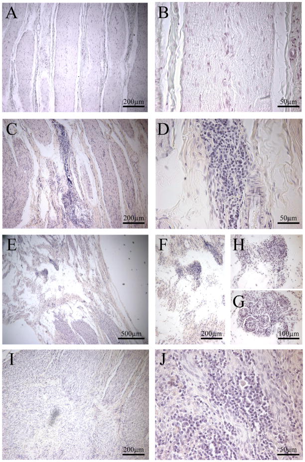
Figure 7.
Exemplars of nerves with injury and inflammation post-surgery. A-B are images of an untreated nerve, with no injury or inflammation. C-J are examples of varying degrees of injury and inflammation apparent in chronic constriction injured nerves taken 2–7 weeks post-surgery. A, C, E, I (left panels) are lower magnification to illustrate nerve injury, as indicated by the infiltration of chronic inflammatory cells, lymphocytes. B,D, F-H, J (right panels) are higher magnification to show the degree of inflammation. G depicts giant cells/foreign body reaction and inflammatory cells. All nerves were stained with hematoxylin and eosin.

