Fig. 1.
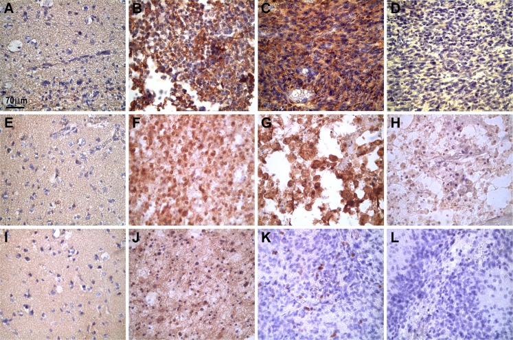
Representative photographs showing the microscopic expression of Hsp27, Hsp70total (recognized by monoclonal antibody BRM22), and Hsp70Ind in normal and tumor biopsy samples. In “normal” brain tissues, the three Hsps were practically absent (a, e, and i). In contrast, in tumor cells, Hsp27 was expressed in the cytoplasm of grade II oligodendroglioma (b) and in grade IV astrocytoma (c). Hsp70total was expressed in the nuclei and cytoplasm of grade II oligodendrogliomas (f) and in grade IV astrocytomas (g). In oligodendrogliomas (grade II) (j), Hsp70Ind was expressed with weak immunostaining intensity in some nuclei and in the background while, in grade IV astrocytomas (k), the Hsp70Ind protein appeared in a few nuclei. Negative controls using isotype antibodies are shown in d, h, and l (grade IV astrocytomas). The images were captured with a Nikon Eclipse E200 microscope (×40 objective). The positive immunoreaction appears as brown deposits; the slides were lightly counterstained with hematoxylin to reveal nuclei

