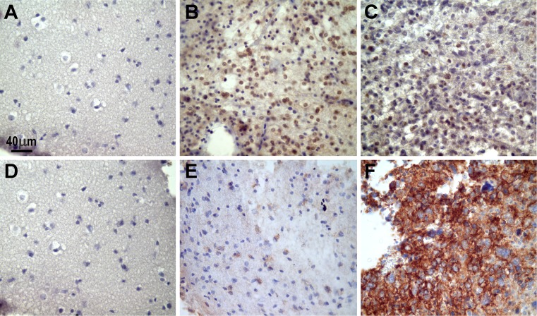
Fig. 4.
Representative photographs of the microscopic expression of MGMT and β-catenin in normal and tumor biopsy samples. “Normal” brain tissues did not show positive immunoreaction for MGMT (a) or β-catenin (d). In contrast, MGMT was noted mainly in the nuclei of oligodendroglioma cells (b), and the expression decreased in this grade II astrocytoma (c). β-catenin appeared as granular deposits in the cytoplasm in this grade II astrocytoma (e). This protein notably increased in a grade IV astrocytoma (note the cytoplasmic immunostaining) (f). The images were captured with a Nikon Eclipse E200 microscope (×40 objective). The positive immunoreaction appears as brown deposits; the slides were lightly counterstained with hematoxylin to reveal nuclei

