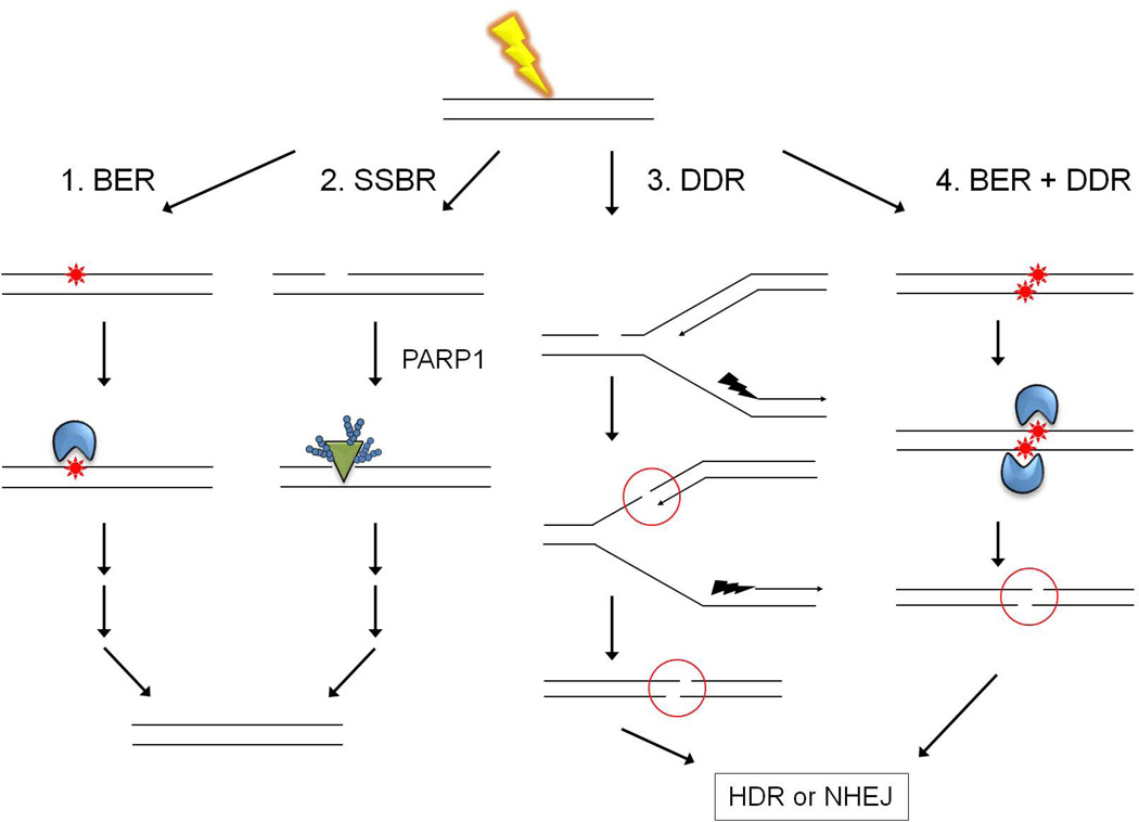
Figure 2. Channeling of oxidatively damaged DNA into multiple DNA repair pathways.
Reactive oxygen species (ROS), in this case generated by exposure to γ-radiation (yellow lightning), can produce single and multiple clustered base damages (red stars), and single strand DNA breaks (SSBs). As depicted in the left-most diagram and in Figure 1, most oxidatively damaged bases and AP sites are repaired in an error-free manner by short-patch BER (pathway 1). SSBs generated as intermediates during BER appear to remain bound by DNA glycosylases (blue) or APE until displaced by the next enzyme in the BER pathway, and are thus protected from PARP1 (green triangle) binding. Binding of PARP1 to de novo-generated SSBs channels substrates into the single-strand break repair pathway (pathway 2). Pathway 3 depicts the generation of a double strand DNA break (DSB) via replication up to a SSB. DSBs are also produced during the attempted, near simultaneous BER of closely opposed lesions (pathway 4). In either case, DSBs trigger what is now commonly called the DNA damage response (DDR), which encompasses both homology-directed repair (HDR) and non-homologous end joining (NHEJ) pathways.

