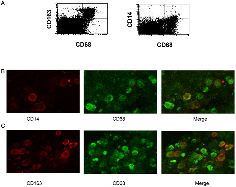
Figure 1. CD163 is highly expressed by uterine macrophages.
Freshly obtained uterine endometrial tissue sections were enzymatically digested into single cell suspensions, stained for CD68 and CD163 or CD68 and CD14 (A) and analyzed by flow cytometry. The numbers inset in the histograms are the percentage of CD163+/CD68+ or CD14+/CD68+ cells. (B and C) Confocal imaging of uterine endometrial macrophages. Endometrial tissues were sectioned and stained for either (B) CD14 and CD68 or (C) CD163 and CD68. Images shown are representative of tissue sections from six individuals.

