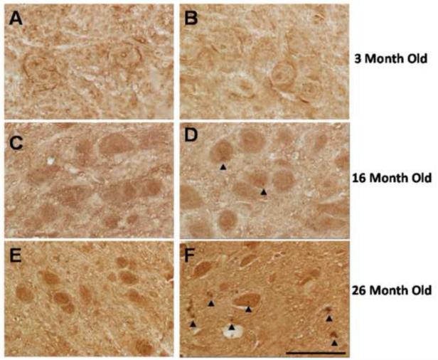
Fig. 8.
Microscopic assessment of age-associated accumulation of ubiquitin-positive intraneuronal inclusions in the WT and OGG1 KO mice. Representative photographs of the substantia nigra of WT (A,C,E) and OGG1 KO (B,D,F) mice. Arrow heads identify intraneuronal ubiquitin aggregates. Scale Bar 50 m.

