
Fig. 1.
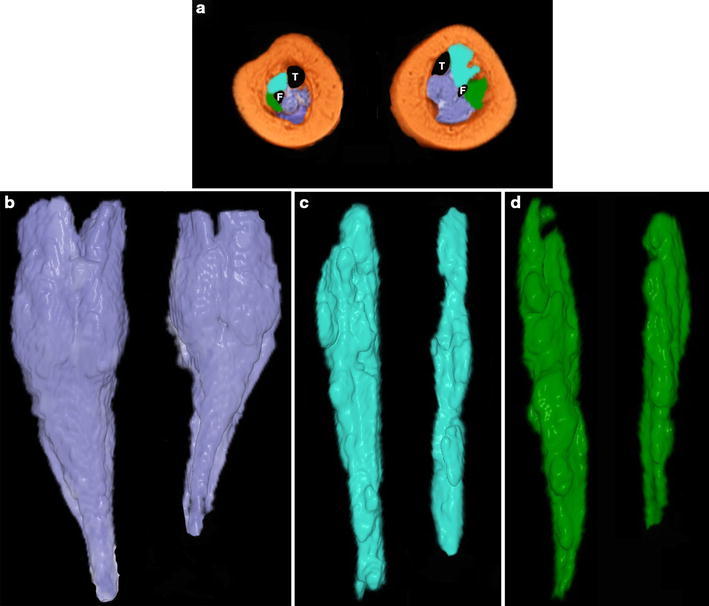
a Magnetic resonance imaging (MRI) cross-section taken at the middle third of the legs in a 4-month-old patient with unilateral congenital right clubfoot (the plantar aspect of the foot faces the reader). The left leg is normal. The postero-medial muscle compartment is coloured in violet, the anterior in sky-blue and the lateral in green. T tibia, F fibula. b The postero-medial compartment is thinner and shorter on the clubfoot side than on the normal side, and the thinnest part starts below the lower end of the gastrocnemius. Both the anterior c and the lateral d muscle compartments are also thinner and shorter than normal on the clubfoot side