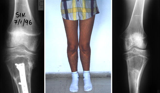
Fig. 7.

Clinical appearance and radiological evaluation 1 year postoperatively of the same patient shown in Fig. 4. X-rays show incomplete correction of the proximal tibia deformity with compensation by the associated distal femoral valgus, but the clinical appearance is more impressive
