
Figure 1.
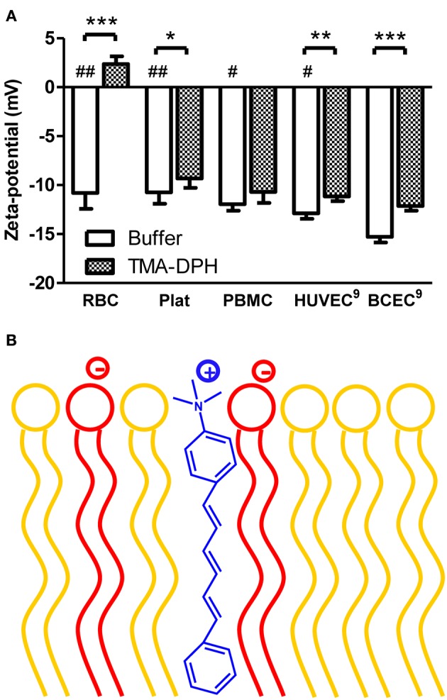
Zeta-potential of blood components and endothelial cells of mammals in the absence and in the presence of TMA-DPH. (A) RBC (4 × 10⁵ cells/mL), platelets (4 × 10⁶ platelets/mL), and PBMC (4 × 10⁵ cells/mL), HUVEC and BCEC (1 × 10⁵ cells/mL) were incubated with TMA-DPH (54 μM) at 25°C and zeta-potential was measured. Data shown as mean ± SEM; each group value is an average of at least two independent measurements. *P < 0.05; **P < 0.01; ***P < 0.001 vs. unlabeled samples, t-test; and #P < 0.05, ##P < 0.01 vs. BCEC, One-Way ANOVA, Bonferroni's multiple comparison test. (B) Schematic representation of TMA-DPH localization in the lipid membrane. The cationic trimethylamino group of TMA-DPH (in blue) locates near the polar heads of phospholipids; anionic phospholipids are represented in red and zwitterionic in yellow.