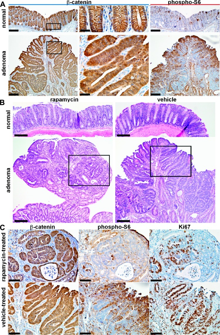
Figure 4.
Rapamycin treatment inhibits mTOR signaling and attenuates cell growth in adenomas with Apc defect. (A) Representative low magnification images show increased total β-catenin expression from adenoma derived from CPC;Apc mice in comparison to normal (left panels; scale bar, 100 µm). Boxed regions are shown in high magnification (middle panels; scale bar, 20 µm). Increased expression of phospho-S6 can be appreciated in adenoma in comparison to normal (right panels; scale bar, 100 µm). (B) Histology (H&E) of normal distal colonic epithelium and adenoma treated with rapamycin (left panels) and vehicle (right panels; scale bars, 100 µm). The boxed areas indicate regions of dysplasia that were further studied on IHC below. (C) Significant levels of total β-catenin expression were observed from the adenomas in both rapamycin and vehicle-treated adenomas (left panels). Level of phospho-S6 ribosomal protein expression is lower in residual adenoma from rapamycin-treated mouse compared to that for the vehicle-treated mouse (middle panels). A significant reduction in the number of Ki67-labeled cells was observed in rapamycin-treated adenomas compared to vehicle-treated adenomas (right panels; scale bars, 25 µm).

