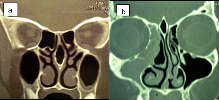
Fig. 1.
Coronal CT scans. a) Nasal septal pneumatization. b) Extensive pneumatization of the crista galli or bulla galli. Right concha bullosa; left pneumatization of the uncinate process; deviation of the nasal septum convexity to the left can be seen; small bilateral Haller cells are also present.

