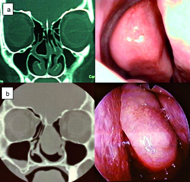
Fig. 2.
Coronal CT scan and corresponding endoscopic image of concha bullosa. a) Large right concha bullosa with moderate deviation of the nasal septum convexity to the left. There is also small Haller cell on the right side, and Keros grade II. b) Masssive left mucopyocele of the concha bullosa of the middle turbinate presenting as a large nasal mass.

