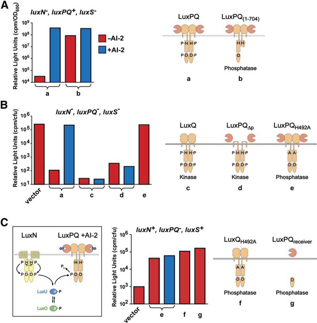
Figure 2. Dimerization and In Vivo Activities of LuxPQ.
Bioluminescence activity was measured in various genetic backgrounds (indicated above each graph); cartoons depict the LuxPQ proteins expressed in trans. The V. harveyi strains used were the following: (A) JMH610 (luxN−, luxPQ+, luxS−), (B) FED119 (luxN−, luxPQ−, luxS−), and (C) BB886 (luxN+, luxPQ−, luxS+). The luxPQ plasmids examined carried the following: (a) wild-type LuxPQ; (b) LuxPQ1–704; (c) LuxQ alone, generated by mutating luxP such that secretion into the periplasm was abolished (LuxPss; see Experimental Procedures); (d) LuxPQΔp (wild-type LuxP, together with LuxQ lacking its periplasmic domain, residues 45–265); (e) LuxPQH492A; (f) LuxQH492A (with an in-frame deletion of luxP); and (g) LuxPQreceiver. V. harveyi AI-2 was generated in situ in the specified cultures (blue bars) by adding 1 µM synthetic DPD in the presence of boric acid. Further details are given in the Experimental Procedures.

